Heart Monitor
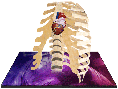
Because the heart is so well protected from outside danger, monitoring the heart can be a challenge. Medical scientists have several ways of "seeing" the heart without actually having to open the chest. Typically, open heart surgery is performed only as the last resort.
To monitor the heart, a doctor begins by touching the chest. If the tip of the heart can be felt pushing against the chest, the heart may be enlarged. Thumping lightly on the chest gives an idea of the heart's shape. Your body's vital statistics also tell how well the heart is working.
Next, the doctor places the stethoscope on the chest to hear the sound of the heart. The normal heart sounds, lub and dub, can be heard. Any unusual sounds can also be heard. Heart arrhythmias and murmurs are two possibilities. An arrhythmia is an irregular pattern of the heart's beat. A murmur indicates that blood is seeping through the closed valve separating the atrium and ventricle.
An x-ray machine passes rays through the chest to make a shadow picture of the heart. The x-ray shows the size and position of the heart. It can also show any obvious deformities.
Modern technology has provided even clearer pictures of the heart. Echocardiography and Electrocardiography are two techniques that provide detailed information about the heart without causing any real discomfort to the patient.
If these procedures do not provide enough information, exploratory procedures may need to be performed. Cardiac catheterization and angioplasty are two common methods for exploring the heart. The last resort, of course, is open heart surgery.