A peek at the secret life of
your heart A peek at the secret life of
your heart
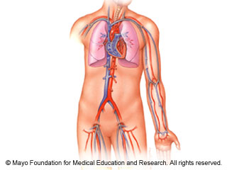
Your heart and a 60,000-mile network of blood
vessels make up your cardiovascular system. Well, it would be that
long if you stretched out all those vessels end to end.
Your heart is responsible for the main function of your
cardiovascular system: pumping blood through this vast network to all
of the tissues and organs of your body, a process called circulation.
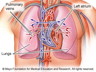
Think of the circulatory process as a figure eight. In one loop,
blood circulates between your heart and lungs (pulmonary circulation).
In the other, blood circulates between your heart and the rest of your
body (systemic circulation). These two loops interact to pump blood in
a continuous circuit: heart to body, body to heart, heart to lungs,
lungs to heart, and heart to body.
How your heart works to pump blood and vital
nutrients throughout your body.
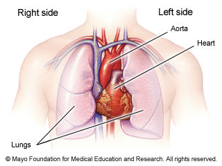
Where is the heart ?
Your heart is a muscular organ located slightly to the left of the
center of your chest — not to the far left, as is sometimes thought.
Your heart isn't quite shaped like a valentine, either. It's
actually shaped like an inverted cone. The tip (apex) is at the
bottom and the wider part at the top. Your heart sits at an angle in
your chest, with the apex pointing to the left.
Your heart is protected by your breastbone (sternum) in the front
and your spinal column in the back, plus your lungs and rib cage.
The average adult heart is about the size of a clenched fist and
weighs about 12 ounces
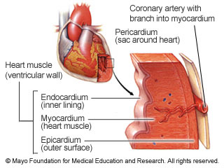
How many layers ?
The muscular wall of the heart has three layers:
Endocardium. The thin inner layer
that covers the inside surfaces of your heart's four chambers,
valves and muscles.
Myocardium. The thick middle
layer of the heart muscle. It's the workhorse, responsible for
most of the heart's pumping action.
Epicardium. The thin, glossy
membrane that covers the outer surface of the heart.
In addition, a protective sac called the pericardium encases
the entire heart
The chambers
Circulation is a continuous process, and your heart,
lungs and the rest of your body have a constant supply of
blood coming and going. Various structures in your heart
regulate those comings and goings.
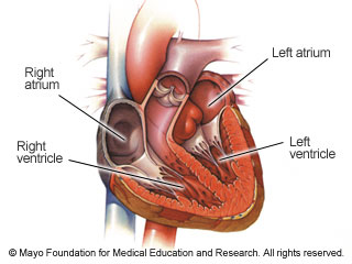
Your heart is divided into two sides, left and right.
Each side has two chambers, or open spaces, for a total of
four. The four chambers are the left atrium and right atrium
near the top of the heart, plus the left ventricle and right
ventricle near the bottom. The right side of the heart is
responsible for pulmonary circulation, while the left side
forms the circulatory loop that supplies blood to the rest
of your body.
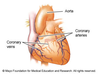
Nourishing the heart
In addition, the heart muscle needs its own supply of blood. Two
branches of the aorta lead to the right and left
coronary arteries. Those arteries extend over the
surface of the heart and branch into smaller
capillaries. The capillaries supply blood to the heart
for its own nourishment. The capillaries drain into two
coronary veins that empty into the right atrium
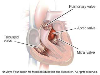
Valves: The heart's gatekeepers
Valves within your heart make sure blood flows in the right direction.
They function much like gates: They open only when
they're pushed on, and they open only one way. The
heart has four valves: the tricuspid, mitral,
pulmonary and aortic. Each opens and closes once per
heartbeat — or about once every second.
The pulmonary circulation loop starts with the
blood sitting in your heart's right atrium. From
that atrium, blood flows through the tricuspid valve
into the right ventricle.
Into the lungs
The tricuspid valve allows blood into the right ventricle. When filled
with blood, the ventricle then contracts, forcing blood
out — much like squeezing ketchup out of a soft bottle.
Contraction, known as systole (SIS-to-le), forces blood
from one area of your heart to another.
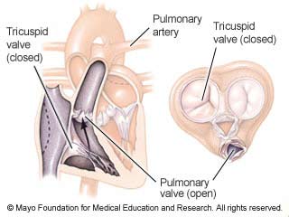
Contraction of the right ventricle forces the
tricuspid valve to close and the pulmonary valve to
open. With the pulmonary valve open, blood can flow into
the pulmonary artery. The pulmonary artery branches into
two vessels, one that carries blood to the left lung and
one that carries blood to the right lung.
Picking up oxygen
Once in the lungs, the blood gives up carbon dioxide gas. The gas
passes through tiny air sacs into bronchioles — tubes
that carry air into and out of the lungs — and you
exhale the carbon dioxide.
When you breathe in, your lungs get a supply of
oxygen. Red blood cells in your blood pick up that
oxygen, turning your blood bright red. The tissues and
organs of your body need oxygen to function, and your
blood carries it to them
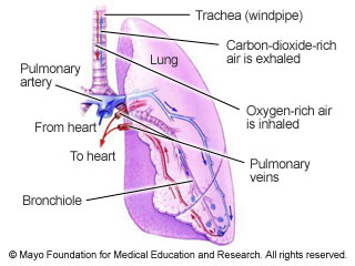
Back to the heart
The blood needs to get out of your lungs so it can distribute that
oxygen. Each lung has two pulmonary veins. The veins
carry that newly oxygenated blood back to your heart,
delivering it to the left atrium, where it can begin
the journey to the rest of your body.
Arteries, veins, capillaries — all of these vessels
may get confusing, but each has its own role. Arteries
carry blood away from the heart. Veins carry blood
back to the heart, whether from the lungs or other
parts of your body, from head to toe. Capillaries are
the go-between, transporting blood from arteries to
veins and through which nutrients enter tissues and
waste products are picked up.
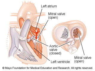
Into the left ventricle
With blood in the left atrium, the heart relaxes — the opposite of
contraction. This phase is called diastole (di-AS-to-le).
The relaxation forces blood from the atrium, pushing
it against the mitral and tricuspid valves — the
gates — which open and allow blood into the left
ventricle.
The cycle of contraction and relaxation causes
blood flow to be pulsatile — it pulsates, or beats
rhythmically. You can often feel your heart beating
by placing your hand on the left side of your chest.
The pulsation is also transmitted to your blood
vessels, so you can feel your pulse where large
arteries are close to the surface of your body, such
as your wrist, your neck and your groin. The period
from the beginning of one heartbeat to the beginning
of the next is the cardiac cycle.
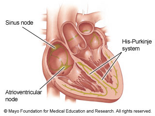
The Electrical system
All this beating of your heart wouldn't be possible, though, without
some electricity. Like your home, your heart has
an electrical (conduction) system. The conduction
system carries electrical impulses throughout your
heart, causing it to beat.
Impulses begin in the sinus node, high in the
right atrium. They travel through the atrial
pathways to the atrioventricular node. There, the
signals briefly slow down as they're funneled into
the electrical network of the ventricles, called
the His-Purkinje (hiz-pur-KIN-je) system. The
conduction system permits the electrical impulse
to reach all parts of your heart at the right
time, so that the heartbeat is coordinated and
occurs at a normal rate. Your heart's electrical
activity can be recorded on an electrocardiogram (ECG).
Into the aorta
With blood in it, the left ventricle then contracts — again beginning
the systolic portion of the cardiac cycle. That
forces the mitral valve to close while opening
the aortic valve. With the aortic valve open,
blood can flow freely into the aorta.
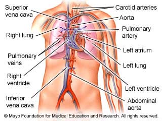
The aorta, or main artery, is the largest
blood vessel in your body. It has numerous
branches to supply various parts of your body
with blood. The branches of the carotid artery,
for instance, travel in your neck to supply your
brain with blood. The aorta also turns downward,
supplying blood vessels to your abdomen. It
splits into more arteries, supplying the legs
with blood.

Starting over again
Your blood transports more than just oxygen and carbon dioxide. It also
carries hormones from endocrine glands, for
instance, making sure they wind up in the right
places. Waste products — other than carbon
dioxide — go to your kidneys and liver, where
they can be removed or broken down. Blood also
picks up nutrients from your intestines and
carries them to your liver and other parts of
your body.
Your tissues and organs absorb oxygen and the
other nutrients from blood. Depleted blood then
flows back to the heart, through a network of
veins, to become replenished. The large veins
that enter the heart are the superior vena cava
and the inferior vena cava. And the circulatory
process starts anew. Actually, there's no
beginning or end to circulation. It's a
continuous and efficient process to supply your
body with all of the nutrients it needs to
function normally — without even thinking about
it.
|
|